Fluoroscopy-guided injection is a medical imaging technique using real-time X-ray visualization to ensure precise needle placement during injections, enhancing accuracy and safety in pain management and orthobiologic therapies.
1.1. Definition and Overview
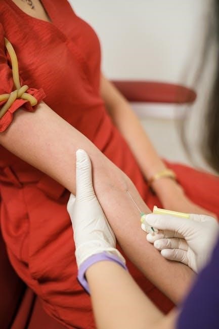
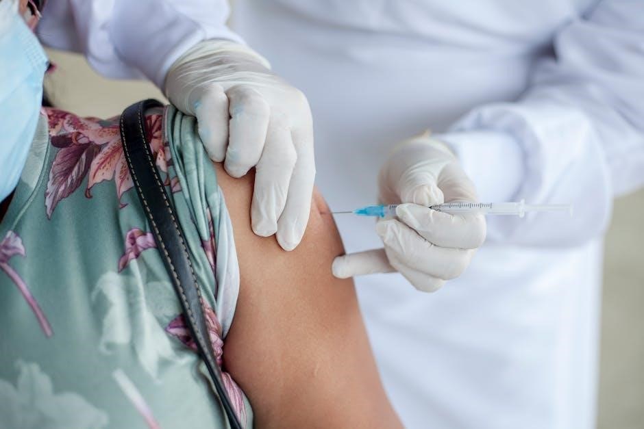
Fluoroscopy-guided injection is a minimally invasive medical procedure that uses real-time X-ray imaging to guide needle placement for precise delivery of medications or therapies. Widely used in pain management and orthobiologic treatments, it enhances accuracy by providing continuous visualization of anatomical structures; This technique is particularly effective for injections targeting specific joints, epidural spaces, or soft tissues. The use of contrast agents further improves visualization, ensuring proper medication distribution. By minimizing blind injection risks, fluoroscopy-guided injection reduces complications and improves therapeutic outcomes, making it a cornerstone in modern interventional medicine.
1.2. Historical Development and Evolution
Fluoroscopy-guided injection traces its roots to the discovery of X-rays by Wilhelm Roentgen in 1895, enabling real-time visualization of internal structures. Early applications focused on diagnostic imaging, with fluoroscopy emerging as a tool for guiding injections in the mid-20th century. Technological advancements, such as image intensifiers and digital fluoroscopy, improved resolution and reduced radiation exposure. Modern systems integrate contrast agents and advanced imaging software, enhancing precision and safety. The technique has evolved into a cornerstone of interventional medicine, offering minimally invasive solutions for pain management, orthobiologic therapies, and other applications, with ongoing innovations continuing to expand its clinical utility;

Fluoroscopy: Basic Principles and Technology

Fluoroscopy is a medical imaging tool using X-rays to create continuous, real-time images of internal structures, functioning like an X-ray movie for diagnostic and procedural guidance.
2.1. How Fluoroscopy Works
Fluoroscopy uses X-rays to produce real-time images of internal structures. An X-ray beam passes through the body, and a phosphor-coated screen emits light, captured by an image intensifier. This intensifies the light, allowing a camera to capture and display continuous images on a monitor. The process enables visualization of dynamic movements, such as needle placement during injections. Contrast agents can enhance visibility of specific areas. This technology provides immediate feedback, making it invaluable for guiding precise medical procedures, ensuring accuracy and safety in interventions like pain management and orthobiologic therapies.
2.2; Radiation Safety and Dose Management
Radiation safety is a critical priority in fluoroscopy-guided injections. Fluoroscopy emits ionizing radiation, posing risks such as tissue damage and increased cancer likelihood at high doses. Minimizing exposure is essential, adhering to the ALARA principle (As Low As Reasonably Achievable). Techniques include using pulse low-dose modes, limiting fluoroscopy time, and shielding non-target areas. Operators monitor dose rates and cumulative exposure. Contrast agents can reduce reliance on high radiation levels. Regular equipment maintenance and staff training further mitigate risks. Balancing diagnostic benefits with radiation risks ensures safe and effective procedures, protecting both patients and medical personnel.
Clinical Applications of Fluoroscopy-Guided Injections
Fluoroscopy-guided injections are widely used in pain management, orthobiologic therapies, and precise drug delivery. Common applications include facet joint injections, epidural procedures, and subarachnoid injections, ensuring accurate targeting of treatment areas.
3.1. Pain Management: Facet Joint and Epidural Injections
Fluoroscopy-guided facet joint and epidural injections are effective treatments for spinal pain. Facet joint injections target inflammation in the small joints of the spine, while epidural injections deliver medication into the epidural space to relieve pain and inflammation. These procedures use real-time imaging to ensure precise needle placement, minimizing complications. They are commonly used for chronic low back pain, radiculopathy, and post-surgical pain. The use of fluoroscopy enhances the accuracy of these injections, leading to better clinical outcomes and patient satisfaction compared to blind or landmark-based techniques.
3.2. Orthobiologic Management of Osteoarthritis
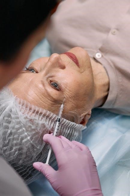
Fluoroscopy-guided injections play a critical role in orthobiologic management of osteoarthritis, enabling precise delivery of regenerative therapies. Orthobiologic agents, such as platelet-rich plasma (PRP), bone marrow aspirate concentrate (BMAC), and hyaluronic acid, are injected into joints or damaged tissues under fluoroscopic guidance. This ensures accurate placement, maximizing therapeutic potential. These treatments aim to promote cartilage regeneration, reduce inflammation, and improve joint function. Fluoroscopy enhances the safety and efficacy of orthobiologic interventions, making them a valuable option for patients seeking minimally invasive, non-surgical solutions for osteoarthritis. Research continues to explore the long-term benefits and optimal applications of these innovative therapies.
3.3. Subarachnoid and Intrathecal Injections
Subarachnoid and intrathecal injections are advanced fluoroscopy-guided procedures used to deliver medications directly into the spinal canal. These injections target the subarachnoid space, where cerebrospinal fluid circulates, or the intrathecal space, for conditions like severe chronic pain or inflammation. Fluoroscopy ensures precise needle placement, minimizing risks of nerve damage or improper drug distribution. Commonly administered medications include local anesthetics, steroids, or other therapeutic agents. These procedures are particularly beneficial for patients with refractory pain or those requiring targeted drug delivery. The use of fluoroscopy enhances both the safety and efficacy of these injections, making them a critical tool in pain management strategies.

Procedure and Technique
Fluoroscopy-guided injection involves a systematic approach, including patient preparation, positioning, and real-time imaging to ensure accurate needle placement and delivery of therapeutic agents.
4.1. Patient Preparation and Positioning
Patient preparation involves reviewing medical history, ensuring NPO status if required, and obtaining informed consent. Positioning is critical, with the patient placed to optimize fluoroscopic visualization of the target anatomy. Comfort and stability are prioritized to minimize movement during the procedure. The use of contrast agents may be planned, and the patient is educated on breathing techniques to enhance image clarity. Proper positioning ensures accurate needle placement, reducing complications and improving procedural success rates.
4.2. Injection Technique and Image Guidance
The injection technique involves advancing the needle under real-time fluoroscopic imaging, allowing precise targeting of the injection site. The operator adjusts the needle trajectory based on fluoroscopic images to ensure accurate placement. Contrast agents are often used to confirm needle position and spread of the injected material. Patient stability is maintained throughout the procedure, with continuous monitoring of the fluoroscopic feed. The technique relies on the operator’s skill in interpreting images and making adjustments to achieve optimal outcomes. This approach minimizes risks and enhances the efficacy of fluoroscopy-guided injections in various clinical applications.
4.3. Use of Contrast Agents
Contrast agents are often employed in fluoroscopy-guided injections to enhance visualization of the injection site and confirm proper needle placement. These agents, typically iodine-based or gadolinium-based, are administered before or during the procedure. They help in delineating soft tissue structures and ensuring the injected material spreads as intended. Real-time imaging allows the operator to verify the contrast agent’s distribution, ensuring accurate delivery. This step is critical for minimizing complications and optimizing therapeutic outcomes. The use of contrast agents is particularly valuable in complex injections, where anatomical precision is paramount. Their application ensures safety and efficacy in fluoroscopy-guided procedures.
Radiation Safety and Biological Effects
Fluoroscopy-guided injections require careful radiation exposure management to minimize biological risks. Operators and staff must adhere to safety guidelines to protect patients and personnel from radiation hazards.
5.1. Radiation Exposure Risks
Radiation exposure during fluoroscopy-guided injections poses risks, including potential biological effects like cancer. Patients and staff are exposed to ionizing radiation, necessitating strict safety protocols. Operators must minimize dose using the ALARA principle. Modern systems reduce exposure through pulsed imaging and lower dose rates. Protective gear, such as lead aprons, is essential for staff. Long-term risks are dose-dependent, emphasizing the need for monitoring and adherence to guidelines. Regulatory bodies like the NCRP oversee safety standards to mitigate these risks, ensuring procedures remain as safe as possible while maintaining diagnostic accuracy.
5.2. Operator and Staff Safety Guidelines
Operator and staff safety is critical during fluoroscopy-guided injections. Protective gear, including lead aprons, thyroid shields, and gloves, is essential to minimize radiation exposure. Personal dosimeters monitor cumulative dose. The ALARA principle—As Low As Reasonably Achievable—guides radiation use. Regular training on radiation safety and proper equipment handling is mandatory. Staff should maintain distance from the radiation source and use shielding when possible. Facilities must implement safety protocols and monitor exposure levels to ensure compliance with regulatory standards, safeguarding both patients and personnel from unnecessary radiation risks while performing fluoroscopy-guided procedures;
Efficacy and Outcomes
Fluoroscopy-guided injections demonstrate high success rates in pain management, with improved accuracy and precision, leading to enhanced patient outcomes and reduced complications compared to blind techniques.
6.1. Fluoroscopy vs. Other Imaging Modalities
Fluoroscopy excels in providing real-time visualization, making it superior for dynamic procedures. Compared to ultrasound, it offers better bone and deep tissue detail, though ultrasound avoids radiation. CT-guidance provides higher image resolution but is less practical for real-time guidance. Fluoroscopy’s balance of accuracy, cost, and accessibility makes it a preferred choice for many injections, despite radiation concerns. Its advantages in guiding complex injections, like epidural and facet joint procedures, are well-documented, ensuring precise needle placement and effective treatment delivery;
6.2. Success Rates and Clinical Evidence
Fluoroscopy-guided injections demonstrate high success rates, particularly in pain management and orthobiologic therapies. Studies show improved accuracy and efficacy compared to blind injections, with reduced complication risks. Clinical evidence supports its use in facet joint and epidural injections, achieving significant pain relief in many patients. However, some studies, like the 2012 Cochrane review, question the added benefit of imaging guidance for certain conditions, such as shoulder injections. Despite this, fluoroscopy remains a gold standard due to its real-time visualization, enhancing precision and therapeutic outcomes. Ongoing research continues to refine its applications and validate its effectiveness across diverse clinical scenarios.
6.3. Limitations and Controversies
Fluoroscopy-guided injections are not without limitations. Some studies, like the 2012 Cochrane review, suggest limited evidence for improved efficacy in certain conditions, such as shoulder pain. Radiation exposure remains a concern, particularly for patients requiring repeated procedures. Additionally, the technique’s success heavily depends on the operator’s skill, introducing variability in outcomes. Cost and accessibility of fluoroscopy equipment can also limit its use in smaller medical facilities. These factors highlight the need for careful patient selection and alternative imaging modalities when appropriate.
Comparison with Other Imaging-Guided Techniques
Fluoroscopy offers real-time imaging, enhancing accuracy, but involves radiation exposure, unlike ultrasound. It is often compared to CT and MRI for precision and invasiveness in guided injections.
7;1. Ultrasound-Guided vs. Fluoroscopy-Guided Injections
Ultrasound-guided injections offer real-time imaging without radiation, making them safer for patients and staff. They are cost-effective and ideal for superficial targets. However, depth penetration is limited, and image quality depends on the operator. Fluoroscopy-guided injections provide superior visualization of deep structures and are highly accurate. They are preferred for complex procedures but involve radiation exposure. Both techniques are effective, with ultrasound being non-invasive and fluoroscopy offering precise guidance. The choice depends on the procedure, patient factors, and clinical context, balancing safety, accuracy, and radiation risks.
7.2. CT-Guided vs. Fluoroscopy-Guided Injections
CT-guided injections provide high-resolution, cross-sectional images, offering exceptional detail for complex anatomical structures. They are ideal for deep tissue targets and precise needle placement. However, CT guidance lacks real-time imaging, requires higher radiation doses, and is less cost-effective. Fluoroscopy-guided injections, while offering real-time visualization, have lower spatial resolution and are better suited for superficial or moving targets. CT is preferred for procedures requiring detailed tissue differentiation, while fluoroscopy excels in dynamic guidance. The choice between the two depends on the clinical scenario, balancing image quality, radiation exposure, and procedural needs.

Future Trends and Advancements
Technological advancements are enhancing fluoroscopy’s image quality and reducing radiation exposure, while expanding its role in regenerative medicine and orthobiologic therapies for improved patient outcomes.
8.1. Technological Innovations in Fluoroscopy
Recent advancements in fluoroscopy include improved image resolution, reduced radiation doses, and enhanced real-time imaging capabilities. Innovations such as 3D reconstruction and AI-driven systems optimize precision and diagnostic accuracy.
8.2. Emerging Applications in Regenerative Medicine
Fluoroscopy-guided injections are increasingly being explored in regenerative medicine for delivering stem cells, platelet-rich plasma, and growth factors. This technique allows precise placement of biologic agents into targeted tissues, enhancing therapeutic outcomes. Real-time imaging ensures accurate needle positioning, minimizing risks and improving efficacy in treatments for conditions like osteoarthritis and tendon injuries. Advances in fluoroscopy are enabling safer and more effective administration of regenerative therapies, offering promising alternatives to traditional surgical interventions.
Fluoroscopy-guided injection represents a cornerstone in modern interventional medicine, offering unparalleled precision and safety for pain management and regenerative therapies. Its real-time imaging capabilities ensure accurate needle placement, reducing complications and improving outcomes. As advancements in technology and biologic therapies continue, fluoroscopy remains a vital tool in addressing complex conditions like osteoarthritis and spinal pain. The integration of regenerative medicine further expands its potential, making it indispensable in contemporary healthcare. With a strong foundation in safety and efficacy, fluoroscopy-guided injection is poised to play an even greater role in advancing medical treatments.