
Fluoroscopy-guided injection is a minimally invasive technique using real-time x-ray imaging for precise administration of medications or anesthetics, aiding in pain management and diagnostic procedures effectively with accuracy․
1․1 Definition and Overview
Fluoroscopy-guided injection is a medical imaging technique that uses real-time x-ray visualization to guide the precise delivery of medications, such as steroids or anesthetics, into targeted tissues․ It allows physicians to monitor the needle placement and medication distribution during procedures, ensuring accuracy and minimizing complications․ Commonly used in pain management, this method is particularly effective for spinal injections, joint injections, and soft tissue interventions․ The real-time imaging capability enhances the efficacy of the treatment while reducing the risk of misplacement․ This approach is widely favored for its ability to provide immediate feedback, making it a cornerstone in interventional radiology and pain medicine․
1․2 Historical Development
The development of fluoroscopy-guided injection traces back to the discovery of X-rays by Wilhelm Conrad Röntgen in 1895․ Early fluoroscopy relied on primitive systems, with physicians using fluorescent screens to visualize X-rays in real-time․ Over the 20th century, advancements in technology led to the integration of television systems in the 1950s, enhancing image quality and accessibility․ The 1980s and 1990s saw fluoroscopy become a cornerstone in interventional pain management, with its application expanding to spinal and joint injections․ Modern systems now incorporate digital imaging and radiation dose management, improving both efficacy and safety․ These historical milestones have shaped fluoroscopy-guided injection into a precise and indispensable tool in contemporary medicine, enabling targeted therapies with minimal invasiveness․
1․3 Importance in Modern Medicine
Fluoroscopy-guided injection holds significant importance in modern medicine due to its ability to enhance precision in administering medications or anesthetics․ It is particularly valuable in pain management, allowing practitioners to target specific anatomical structures accurately, minimizing risks of complications․ The real-time imaging capability ensures that injections are delivered to the intended site, improving therapeutic outcomes․ This technique is widely used in interventional pain procedures, such as spinal and joint injections, where accuracy is critical․ Additionally, fluoroscopy-guided injection aids in diagnosing pain generators and reducing reliance on surgical interventions․ Its effectiveness in both therapeutic and diagnostic contexts makes it an indispensable tool in contemporary medical practice, enhancing patient care and satisfaction․

Principles of Fluoroscopy
Fluoroscopy is a real-time imaging technique using x-rays to create moving images of internal structures․ It involves x-ray beams penetrating tissues, with varying absorption rates generating images, enabling precise guidance for injections․
2․1 Basic Principles of Fluoroscopy
Fluoroscopy operates by transmitting x-ray beams through a patient’s body, capturing images on a fluorescent screen․ The x-rays are absorbed differently by various tissues, creating contrast․ A camera then converts these images into video, displayed in real-time․ This technology allows healthcare providers to visualize dynamic processes, such as joint movements or fluid injections, with high accuracy․ The system includes an x-ray tube, image intensifier, and video processing unit, ensuring clear and detailed imaging․ This real-time visualization is crucial for guiding injections precisely, reducing complications and improving therapeutic outcomes in procedures like spinal and joint injections․

2․2 Radiation Safety and Dose Management
Radiation safety is critical in fluoroscopy-guided injections to minimize exposure for both patients and operators․ The ALARA principle (As Low As Reasonably Achievable) guides dose management, ensuring that radiation levels are kept minimal while maintaining image quality․ Protective measures include lead aprons, thyroid collars, and gloves for operators․ Patients are selected based on clinical necessity, and alternative imaging modalities are considered when appropriate․ Modern fluoroscopy systems feature dose-reduction technologies, such as pulsed fluoroscopy and last-image hold, to limit radiation exposure․ Regular monitoring of radiation doses and adherence to safety protocols are essential to mitigate risks while ensuring effective procedural outcomes․
Fluoroscopy-Guided Injection Procedures
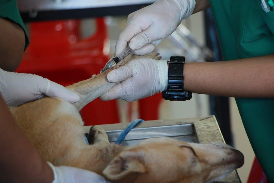
Fluoroscopy-guided injections use real-time x-ray imaging to precisely target tissue, ensuring accurate medication delivery for pain relief, reducing complications, and improving therapeutic outcomes in various clinical settings effectively․
3․1 Preparation for the Procedure

Preparation for fluoroscopy-guided injection involves patient consent, medical history review, and physical assessment․ Patients are instructed to remove metal objects and wear appropriate clothing․ Fasting may be required depending on the procedure․ The injection site is cleaned and sterilized, and local anesthesia is often administered to minimize discomfort․ Equipment, including the fluoroscope and sterile needles, is readied․ The patient is positioned to optimize fluoroscopic visualization․ Vital signs are monitored, and sedation may be provided for anxious patients․ Clear communication ensures patient understanding and cooperation, enhancing safety and procedural success․
3․2 Patient Selection and Education
Patient selection for fluoroscopy-guided injection involves evaluating medical history, current symptoms, and suitability for the procedure․ Education is crucial to ensure understanding and cooperation․ Patients are informed about the procedure’s purpose, benefits, risks, and alternatives․ They are also advised on preparation steps, such as fasting or medication adjustments․ Written and verbal consent are obtained, ensuring patients are aware of potential complications․ Additionally, patients are educated on post-procedure care, including activity restrictions and signs of complications to monitor․ Clear communication helps alleviate anxiety and ensures patients are active participants in their care․ Proper education enhances safety, compliance, and overall procedural outcomes, making it a cornerstone of effective patient management․
3․3 Injection Technique and Positioning
Fluoroscopy-guided injection technique requires precise positioning and real-time imaging to ensure accuracy․ Patients are positioned to optimize access to the target area, such as lying prone or lateral decubitus․ The skin is prepped with antiseptic, and local anesthesia may be used to minimize discomfort․ Under fluoroscopic guidance, the needle is advanced to the desired location, with continuous imaging confirming proper placement․ Contrast agents are often used to verify the injection site․ Proper positioning and technique are critical to avoid complications and ensure therapeutic efficacy․ The procedure is performed under sterile conditions to reduce infection risks․ Real-time imaging allows adjustments, enhancing safety and precision throughout the process․
3․4 Real-Time Imaging Guidance
Real-time imaging guidance is a cornerstone of fluoroscopy-guided injections, enabling precise visualization of the needle and target tissue during the procedure․ Fluoroscopy provides dynamic, high-resolution images, allowing immediate adjustments to needle placement․ This capability minimizes the risk of misplacement and enhances the accuracy of medication delivery․ The use of contrast agents further improves visualization, confirming the correct dispersion of the injected material․ Continuous monitoring ensures that the procedure is performed safely and effectively, reducing complications․ Real-time feedback is essential for achieving optimal therapeutic outcomes, making fluoroscopy an invaluable tool in interventional procedures․ The technology supports both diagnostic and therapeutic interventions with exceptional clarity and control․
Applications of Fluoroscopy-Guided Injections
Fluoroscopy-guided injections are widely used in spinal, joint, and soft tissue procedures, offering precise delivery of medications for pain relief and diagnostic purposes with high accuracy and minimal invasiveness․
4․1 Spinal Injections
Fluoroscopy-guided spinal injections are commonly used to manage chronic pain and diagnose spinal conditions․ These procedures often involve delivering steroids or anesthetics into the epidural space, facet joints, or nerve roots․ Real-time imaging ensures precise needle placement, reducing complications․ Spinal injections are particularly effective for patients with herniated discs, spinal stenosis, or spondylolisthesis․ Fluoroscopy allows visualization of the needle trajectory, enhancing accuracy and safety․ This technique is also used to confirm the pain generator before more invasive treatments․ By providing targeted relief, fluoroscopy-guided spinal injections delay or avoid the need for surgery, offering a minimally invasive solution for pain management․
4․2 Joint and Soft Tissue Injections
Fluoroscopy-guided joint and soft tissue injections are essential for treating inflammatory and degenerative conditions like arthritis, tendinitis, and bursitis․ These injections deliver therapeutic agents, such as corticosteroids or hyaluronic acid, directly into the affected joint or soft tissue․ Fluoroscopy ensures precise needle placement, reducing the risk of misplacement and improving therapeutic outcomes․ Common applications include injections in the shoulder, hip, and knee joints․ Real-time imaging allows for accurate targeting of the inflamed area, minimizing procedural complications․ This technique is particularly beneficial for patients with limited mobility or complex anatomy․ By enhancing the delivery of medications, fluoroscopy-guided injections provide effective relief from pain and inflammation in joints and surrounding soft tissues․
4․3 Musculoskeletal Applications
Fluoroscopy-guided injections are widely used in musculoskeletal medicine to treat conditions like tendinopathy, ligament sprains, and muscle strains․ These procedures often involve injecting corticosteroids, anesthetics, or platelet-rich plasma into affected areas․ Real-time imaging ensures precise needle placement, enhancing the accuracy of therapy delivery․ Common applications include injections for rotator cuff injuries, hip flexor strains, and plantar fasciitis․ Fluoroscopy allows visualization of needle placement relative to tendons, ligaments, and muscles, minimizing complications․ This technique is particularly valuable for deep tissue injections, where anatomical landmarks are less accessible․ By providing immediate feedback, fluoroscopy-guided injections optimize therapeutic outcomes and reduce the risk of injection-related complications in musculoskeletal disorders․

Comparison with Other Imaging Modalities
Fluoroscopy-guided injection is compared to ultrasound and CT-guided techniques, each offering unique advantages․ Fluoroscopy provides real-time imaging, ideal for dynamic procedures, while ultrasound is radiation-free but lacks depth penetration․ CT offers high-resolution images but is less practical for real-time guidance, making fluoroscopy a balanced choice for musculoskeletal and spinal interventions․
5․1 Ultrasound-Guided vs․ Fluoroscopy-Guided Injections
Ultrasound-guided and fluoroscopy-guided injections are both widely used for precise medication delivery but differ in key aspects․ Ultrasound offers real-time imaging without radiation, making it safer for superficial tissues and reducing long-term radiation risks․ However, it may lack the depth penetration needed for complex spinal or deep musculoskeletal injections․ Fluoroscopy provides superior visualization of bony structures and is ideal for dynamic procedures, though it involves radiation exposure․ The choice between modalities depends on the procedure’s requirements, patient factors, and operator expertise․ Both methods aim for accuracy and efficacy, but fluoroscopy remains the gold standard for interventions requiring detailed, real-time bone and soft tissue imaging․ Each has its advantages and limitations, guiding clinical decision-making․
5․2 CT-Guided vs․ Fluoroscopy-Guided Injections
CT-guided and fluoroscopy-guided injections differ in imaging capabilities and clinical applications․ CT provides high-resolution, cross-sectional images, offering superior detail for complex anatomical structures, particularly in deep tissues․ However, it lacks real-time guidance, making it less suitable for dynamic procedures․ Fluoroscopy, with its real-time imaging, excels in guiding injections during movement, such as spinal procedures, but has lower resolution compared to CT․ Radiation exposure varies, with CT often delivering higher doses for detailed imaging, while fluoroscopy’s dose depends on procedure length․ CT is ideal for precise targeting in challenging cases, whereas fluoroscopy is preferred for its cost-effectiveness and availability․ Both modalities coexist, chosen based on procedural needs, patient factors, and desired outcomes․

Potential Complications and Risks
Fluoroscopy-guided injections may pose risks, including bleeding, infection, nerve damage, and allergic reactions․ Radiation exposure is a concern, emphasizing the need for dose management and safety protocols to minimize risks effectively․
6․1 Common Complications
Common complications of fluoroscopy-guided injections include bleeding, hematoma, or infection at the injection site․ Nerve injury or irritation may occur, leading to temporary or, rarely, permanent neurological deficits․ Allergic reactions to the injected medication or contrast agents are possible, ranging from mild skin rashes to life-threatening anaphylaxis․ Radiation exposure, though minimized, carries long-term risks such as cancer, particularly with cumulative doses․ Additionally, unintended injection into blood vessels or surrounding tissues can cause systemic effects or reduced procedural efficacy․ Proper patient selection, sterile technique, and real-time imaging guidance are critical to mitigating these risks and ensuring safe outcomes․
6․2 Radiation Exposure Risks
Radiation exposure during fluoroscopy-guided injections poses potential risks, including increased cancer risk and genetic mutations․ Prolonged or high-dose exposure can lead to biological effects, though modern systems minimize doses․ Patients with cumulative exposure or pregnancy are at higher risk․ Operators and staff must adhere to strict safety protocols, such as using lead shielding and the ALARA principle (As Low As Reasonably Achievable)․ Regular dose monitoring and training are essential to ensure safety․ Patients should discuss personal risk factors with their provider to weigh benefits against potential radiation risks․ Advanced equipment and techniques further reduce exposure, enhancing safety for both patients and medical teams during procedures․
6․3 Minimizing Risks and Complications
To minimize risks and complications in fluoroscopy-guided injections, proper patient selection and adherence to radiation safety protocols are essential․ Using modern fluoroscopy systems with dose-reduction features helps lower radiation exposure․ Operators should employ the ALARA principle, ensuring the lowest necessary dose for procedure success․ Patient education on risks and benefits is crucial․ Additionally, precise injection techniques and real-time imaging guidance reduce complications․ Post-procedure monitoring for adverse effects, such as bleeding or infection, is recommended․ Regular training for healthcare providers and maintaining equipment quality further enhance safety․ By combining these strategies, the likelihood of complications is significantly reduced, optimizing outcomes for patients undergoing fluoroscopy-guided injections․
Future Trends and Advances
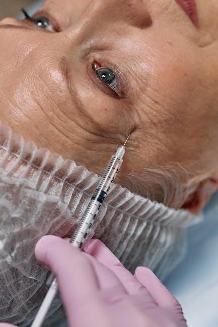
Advancements in fluoroscopy-guided injections include enhanced imaging technologies, reducing radiation exposure and improving precision, alongside AI integration for real-time guidance and personalized treatment approaches, ensuring safer procedures․
7․1 Emerging Technologies in Fluoroscopy
Emerging technologies in fluoroscopy include advanced imaging systems with higher resolution and lower radiation doses․ AI-driven algorithms enhance image clarity and assist in real-time guidance, improving precision․ Portable fluoroscopy units are gaining traction, offering flexibility in various clinical settings․ Additionally, 3D reconstruction capabilities integrated with fluoroscopy provide comprehensive views, aiding in complex injections․ Innovations like dose-reduction systems minimize radiation exposure while maintaining image quality․ These advancements aim to optimize patient safety, improve diagnostic accuracy, and expand the range of fluoroscopy-guided procedures, making them more accessible and effective in modern medical practice․
7․2 Enhanced Imaging Capabilities
Fluoroscopy-guided injection benefits from enhanced imaging capabilities, including high-resolution displays and advanced software for clearer visualization․ Real-time imaging allows precise needle placement, reducing procedural complications․ New systems feature higher frame rates and improved contrast, enhancing anatomical detail․ AI integration aids in noise reduction and automatic brightness adjustments, optimizing image quality․ These advancements enable better visualization of small structures and enhance the accuracy of injections․ Enhanced imaging also supports lower radiation doses while maintaining clarity, improving patient safety․ Additionally, tools like zoom and pan functionalities allow practitioners to focus on specific areas, making procedures more efficient and reducing the need for repeat injections․ These capabilities collectively improve patient outcomes and procedural success rates significantly․
Fluoroscopy-guided injection is a precise and effective technique for accurate medication delivery, emphasizing adherence to guidelines, proper training, and radiation safety to optimize patient outcomes and minimize risks․
8․1 Summary of Key Points
Fluoroscopy-guided injection is a highly precise imaging technique enabling real-time visualization for accurate medication delivery, essential for pain management, diagnostic clarity, and minimally invasive procedures․ It combines x-ray technology with contrast agents to enhance visualization of target structures, ensuring safe and effective treatment outcomes․ Proper patient selection, thorough preparation, and adherence to radiation safety protocols are critical to minimizing risks․ Regular training for operators and continuous advancements in technology further enhance the efficacy and safety of these procedures, making them indispensable in modern medicine for addressing spinal, joint, and musculoskeletal conditions effectively․ Best practices emphasize radiation dose management and personalized patient care to optimize results while reducing complications․
8․2 Guidelines for Effective Use
To ensure the safe and effective use of fluoroscopy-guided injections, proper training and adherence to radiation safety protocols are essential․ Operators should follow the ALARA principle (As Low As Reasonably Achievable) to minimize radiation exposure․ Patient selection and education are critical, with clear communication about risks, benefits, and post-procedure care․ Real-time imaging should be used judiciously, focusing only on the target area․ Documentation of the procedure, including dosage and technique, is vital for accountability and future reference․ Regular maintenance of equipment and quality improvement initiatives further enhance safety and efficacy․ By adhering to these guidelines, healthcare providers can optimize outcomes while minimizing complications, ensuring responsible use of fluoroscopy-guided injections in clinical practice․